Breast Revision Status Post Previous Fat Grafting And Fat Necrosis Of Bilateral Breast
Posted On: June 23, 2016 Author: The Office of Dr. Stuart Linder Posted In: Breast Implants, Breast Reconstruction, Breast Revision, Fat Grafting
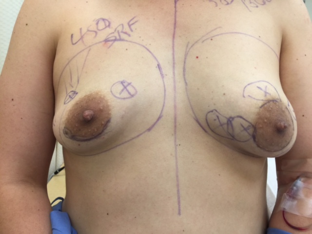
Pre Op Photo
This is the case study of a patient that presents two years post-autologous fat grafting from a different surgeon. Instead of undergoing augmentation mammoplasty using saline or silicone implants, a different doctor decided to do fat grafting of her breasts. Over the last two years she has developed very large cystic fat masses in both breasts, smaller on the right and up to 3 x 6 cm on the left. The patient underwent a mammogram and ultrasound showing enlarging fat cystic masses, especially in the left breast. Five extend from the medial aspect of the breast to the lateral, to the superior 12 o’clock position and to the 3 o’clock position. They are palpable with the largest one almost the size of a golf ball along the lateral left breast.
The patient underwent surgical reconstruction and surgical excisional biopsies of these multiple masses as well as reconstruction using silicone gel implants in order to regain symmetry and correct the deformity of the left breast after removal of these large cystic masses.
In the operating room the patient was placed under general anesthesia. The right breast was first operated upon. A 450 cc SRF silicone gel implant
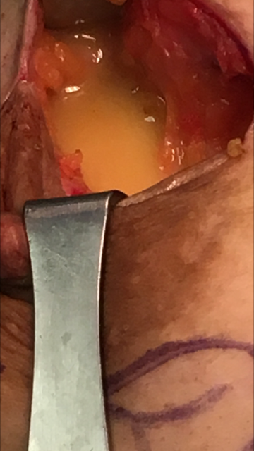 was placed through the periareolar and subpectoral dual plane techniques. The left breast was then incised under the left areola at which time cystic masses were removed along the left medial two large masses and upon identifying fat necrotic liquified tissue was identified. (See photograph to the right) After removing the cystic capsule, the area was coagulated with electrocautery. All cystic masses were then opened along the superior 12 o’clock position, 3 o’clock and then the largest along the left lateral breast. All of them had liquified fat within them, yellow and thick viscous fat in liquified form and the capsules were all exenterated, removed and then bovied. A 385 cc SRF gel was placed on the left to regain symmetry and the patient will be maintained on oral antibiotics for 14 days. A Dr. Linder Bra and upper pole compression band with sutures to remain in place for 14 to 17 days.
This is an interesting case study, showing that fat grafting does not always work to the breast. Not only is there the possibility for misdiagnosis of tumors, but the fat may not survive, leaving the patient with enlarging cystic fat tumors or fat masses which should be surgically removed.
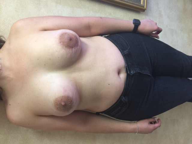
Six Weeks Post Op
The photo to the left shows the patient after six weeks post op, and she is very please with her breast revision.
Schedule your consultation with Dr. Linder to learn more about breast revision surgery. Call our office at (310) 275-4513 in Beverly Hills or fill out our online contact form today.